Home
Uncategories
Anatomy Of Chest : Amazon Com Anatomy Chest Artery Rib Print Sra3 12x18 Conqueror Laid Paper Handmade - This page provides an overview of the chest muscle group.
Anatomy Of Chest : Amazon Com Anatomy Chest Artery Rib Print Sra3 12x18 Conqueror Laid Paper Handmade - This page provides an overview of the chest muscle group.
Anatomy Of Chest : Amazon Com Anatomy Chest Artery Rib Print Sra3 12x18 Conqueror Laid Paper Handmade - This page provides an overview of the chest muscle group.. The dominant muscle in the upper chest is the pectoralis major. The sternum, or breastbone, is a flat bone at the front center of the chest. Anatomy of the eye 12 photos of the anatomy of the eye anatomy of the eye bones, anatomy of the eye lacrimal gland, anatomy of the eye special senses vision, anatomy of the eye ultrasound, external anatomy of the eye quiz, human anatomy, anatomy of the eye bones, anatomy of the eye lacrimal gland, anatomy … Chest bone, ribs, lung, heart, xiphoid process, sternum anatomy. This atlas is a comprehensive and affordable learning tool for medical students and residents and especially for radiologists and pneumologists.
Skandalakis chest wall embryogenesis the muscles of the chest develop from the somites found in the mesoderm. Radiology basics of chest ct anatomy with annotated coronal images and scrollable axial images to help medical students and junior doctors learning anatomy. The chest wall is a complex system that provides rigid protection to the vital organs such as the heart, lungs, and liver; Related posts of anatomy of the chest and stomach anatomy of the eye. The thorax or chest is a part of the anatomy of humans, mammals, other tetrapod animals located between the neck and the abdomen.
Chest Anatomy Stock Photos Offset from ak.picdn.net It provides access to ct images in the axial plane, allowing the user to learn and review the lung anatomy interactively. This chapter is an abbreviated review of thoracic anatomy as seen on chest radiographs and computed tomography (ct) of the chest. The breast is the tissue overlying the chest (pectoral) muscles. Here, we break down the anatomy of your chest muscles. Radiology basics of chest ct anatomy with annotated coronal images and scrollable axial images to help medical students and junior doctors learning anatomy. The chest is the area of origin for many of the body's systems as it houses organs such as the heart, esophagus, trachea, lungs, and thoracic diaphragm. 12 cm (5 in) in length, 8 cm (3.5 in) wide, and 6 cm (2.5 in) in thickness. Anatomically, the heart is located in the anterior thoracic cavity;
The chest or thorax is the region between the neck and diaphragm that encloses organs, such as the heart, lungs, esophagus, trachea, and thoracic diaphragm.
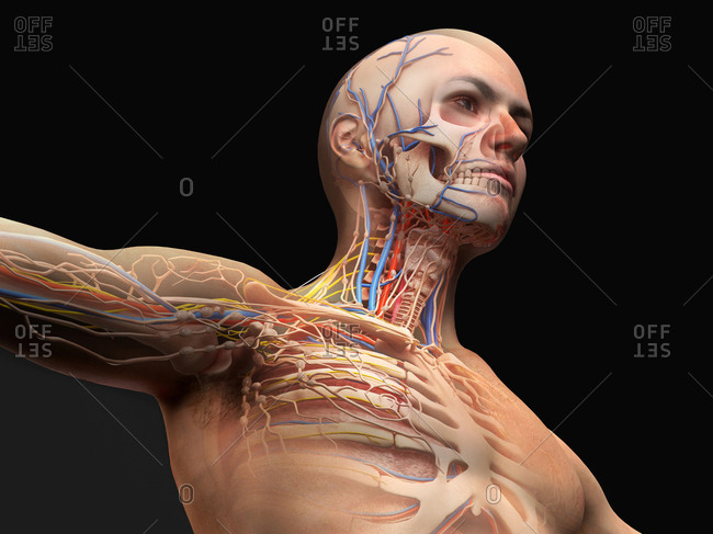
Anatomy of the eye 12 photos of the anatomy of the eye anatomy of the eye bones, anatomy of the eye lacrimal gland, anatomy of the eye special senses vision, anatomy of the eye ultrasound, external anatomy of the eye quiz, human anatomy, anatomy of the eye bones, anatomy of the eye lacrimal gland, anatomy … Swensen fund for innovation in teaching. Anatomy of the thorax, heart, abdomen and pelvis recommended text gray's anatomy for students. However, the classical anatomical descriptions in textbooks make it difficult to gain full mastery of this subject, because the books usually deal with its elements separately. Plus, how to target each to make them bigger and stronger. The circulatory system does most of its work. Summary:for adequate treatment of patients with breast cancer, mastologists should have a complete understanding of the anatomy of the thoracic wall, axilla and breast. Sternocleidomastoid muscle clavicle and ribs anatomy muscle anatomy chest sternocleidomastoid ribs anatomy chest muscles anatomy thorax rib muscles chest muscles chest anatomy illustration. Anatomically, the heart is located in the anterior thoracic cavity; This tutorial is designed to help you understand the normal anatomy of the chest as seen on ct images in three planes: Related posts of anatomy of the chest and stomach anatomy of the eye. The pec major) is the one that commands the most real estate. Chest bone, ribs, lung, heart, xiphoid process, sternum anatomy.
It's also sometimes referred to as the breastbone. Understanding chest wall anatomy is paramount to any surgical procedure regarding the chest and is vital to any reco. Skandalakis chest wall embryogenesis the muscles of the chest develop from the somites found in the mesoderm. Anatomy of the eye 12 photos of the anatomy of the eye anatomy of the eye bones, anatomy of the eye lacrimal gland, anatomy of the eye special senses vision, anatomy of the eye ultrasound, external anatomy of the eye quiz, human anatomy, anatomy of the eye bones, anatomy of the eye lacrimal gland, anatomy … This atlas is a comprehensive and affordable learning tool for medical students and residents and especially for radiologists and pneumologists.
Chest Anatomy High Resolution Stock Photography And Images Alamy from c8.alamy.com Applied anatomy of the chest wall and mediastinum petros mirilas michael e. Chest a man's chest — like the rest of his body — is covered with skin that has two layers. Swensen fund for innovation in teaching. See human chest anatomy stock video clips. Learn about each of these muscles, their locations, functional anatomy and exercises for them. Of the two chest muscles, the pectoralis major (a.k.a. Anatomy of rib cage 12 photos of the anatomy of rib cage anatomical rib cage necklace, anatomy and. Here, we break down the anatomy of your chest muscles.
The pec major) is the one that commands the most real estate. This atlas is a comprehensive and affordable learning tool for medical students and residents and especially for radiologists and pneumologists. Applied anatomy of the chest wall and mediastinum petros mirilas michael e. Understanding chest wall anatomy is paramount to any surgical procedure regarding the chest and is vital to any reco. Your sternum is a bone that's located in the middle of your chest. A good radiologist knows the anatomy because knowing where structures normally live and recognizing the location of an abnormality helps to make or narrow the differential diagnosis. How to view the anatomical labels. The circulatory system does most of its work. A typical heart is approximately the size of your fist: This chapter is an abbreviated review of thoracic anatomy as seen on chest radiographs and computed tomography (ct) of the chest. The dominant muscle in the upper chest is the pectoralis major. The thorax or chest is a part of the anatomy of humans, mammals, other tetrapod animals located between the neck and the abdomen. Stability to arm and shoulder movement;
Diseases of the chest and chest abnormalities make up a significant portion of a physician's daily practice. Radiology basics of chest ct anatomy with annotated coronal images and scrollable axial images to help medical students and junior doctors learning anatomy. Of the two chest muscles, the pectoralis major (a.k.a. How to view the anatomical labels. Here, we break down the anatomy of your chest muscles.
Anatomy Study Chest Muscles By Dipnusurf On Deviantart from images-wixmp-ed30a86b8c4ca887773594c2.wixmp.com This chapter is an abbreviated review of thoracic anatomy as seen on chest radiographs and computed tomography (ct) of the chest. Chest a man's chest — like the rest of his body — is covered with skin that has two layers. Stability to arm and shoulder movement; However, the classical anatomical descriptions in textbooks make it difficult to gain full mastery of this subject, because the books usually deal with its elements separately. Your sternum is a bone that's located in the middle of your chest. Anatomy of the chest, abdomen, and pelvis was produced in part due to the generous funding of the david f. (1) the pectoralis major, and (2) the pectoralis minor. Summary:for adequate treatment of patients with breast cancer, mastologists should have a complete understanding of the anatomy of the thoracic wall, axilla and breast.
Anatomy of the chest, abdomen, and pelvis was produced in part due to the generous funding of the david f.
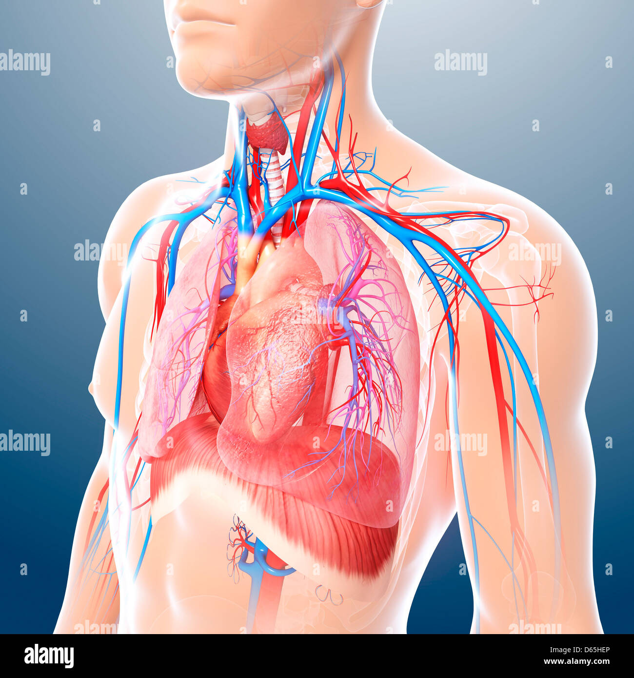
It is enclosed by the ribs, the vertebral column, and the sternum, or breastbone, and is separated from the abdominal cavity (the body's largest hollow space) by a muscular and membranous partition, the diaphragm. It provides access to ct images in the axial plane, allowing the user to learn and review the lung anatomy interactively. And flexibility to aid in the functional process of respiration. This atlas is a comprehensive and affordable learning tool for medical students and residents and especially for radiologists and pneumologists. Anatomy of the thorax, heart, abdomen and pelvis recommended text gray's anatomy for students. Chest a man's chest — like the rest of his body — is covered with skin that has two layers. This chapter is an abbreviated review of thoracic anatomy as seen on chest radiographs and computed tomography (ct) of the chest. The circulatory system does most of its work. Stability to arm and shoulder movement; In this image, you will find common carotid arteries, internal jugular vein, subclavian artery, subclavian vein, heart, right lung, 6th rib, diaphragm, costal cartilage in it. The epidermis is the outermost layer that provides a protective, waterproof seal over the body. Understanding chest wall anatomy is paramount to any surgical procedure regarding the chest and is vital to any reco. A good radiologist knows the anatomy because knowing where structures normally live and recognizing the location of an abnormality helps to make or narrow the differential diagnosis.

0 Comments:
Post a Comment